Female Upper Thigh Anatomy - Thigh Muscles | Anatomy | Pinterest | Sculpture, Google ... / 12 photos of the muscle anatomy of upper thigh.. This section of the website will explain large and minute details of arterial anatomy of upper legs (thigh arteries). Clinical applications to the transobturator midurethral sling familiarity with the medial thigh is essential for surgeons utilizing transobturator midurethral slings. Gluteal tuberosity and upper 1/4 of linea aspera. Quadriceps tendon into patella, then via ligamentum patellae into tubercle of tiba. Learn vocabulary, terms and more with flashcards, games and other study tools.
This webpage presents the anatomical structures found on thigh mri. Gluteal tuberosity and upper 1/4 of linea aspera. • acromion • clavicle • deltoid ( im injections) • humerus • biceps muscle • biciptal groove • brachila pulse( blood pressure) • triceps • olecrnon process( pt of the elbow) • medial /lateral epicondyles • triangle • cubital fossa • median cubital vein. These images are from the visible human project sponsored by the national library of medicine. The nerves of the upper limb arise from a complex arrangement of nerve fibers known as the brachial plexus;
Lower Extremity from classroom.sdmesa.edu In the upper thigh two distinct groups of superficial collectors were found. Learn about the placement of the skeletal and muscular structures. Deviantart is the world's largest online social community for artists and art enthusiasts. To practice tricky questions and answers on all areas of human anatomy, here is complete set of 1000+ multiple choice questions and answers. Want to learn more about it? Upper limb anatomy arm anatomy muscle anatomy anatomy study body anatomy anatomy thigh: 252 360 просмотров • 12 февр. Origin:upper part of intertrochanteric line.
Anatomy atlases, the anatomy atlases logo, and a digital library of anatomy information are all trademarks of michael p.
Collection by renaud galand • last updated 12 weeks ago. In human anatomy, the thigh is the area between the hip (pelvis) and the knee. Anyway, here r some anatomy practices for cheshire 12 photos of the muscle anatomy of upper thigh. The single bone in the thigh region is called the femur. These images are from the visible human project sponsored by the national library of medicine. There may be variations in treatment that. • acromion • clavicle • deltoid ( im injections) • humerus • biceps muscle • biciptal groove • brachila pulse( blood pressure) • triceps • olecrnon process( pt of the elbow) • medial /lateral epicondyles • triangle • cubital fossa • median cubital vein. These nerves give sensation to our upper limb, as well as innervating the muscles, allowing us to move them at will. Want to learn more about it? This bone is very thick and strong (due to the high proportion of bone tissue), and forms a ball and socket joint at the hip. 252 360 просмотров • 12 февр. In the upper thigh two distinct groups of superficial collectors were found. .female pelvis anatomy ct thigh anatomy mri pelvic mri anatomy sciatic nerve anatomy ct anatomy calf muscle anatomy mri thigh musculature ct anterior thigh muscles anatomy anatomy thigh compartments anatomy leg artery anatomy upper leg anatomy sartorius.
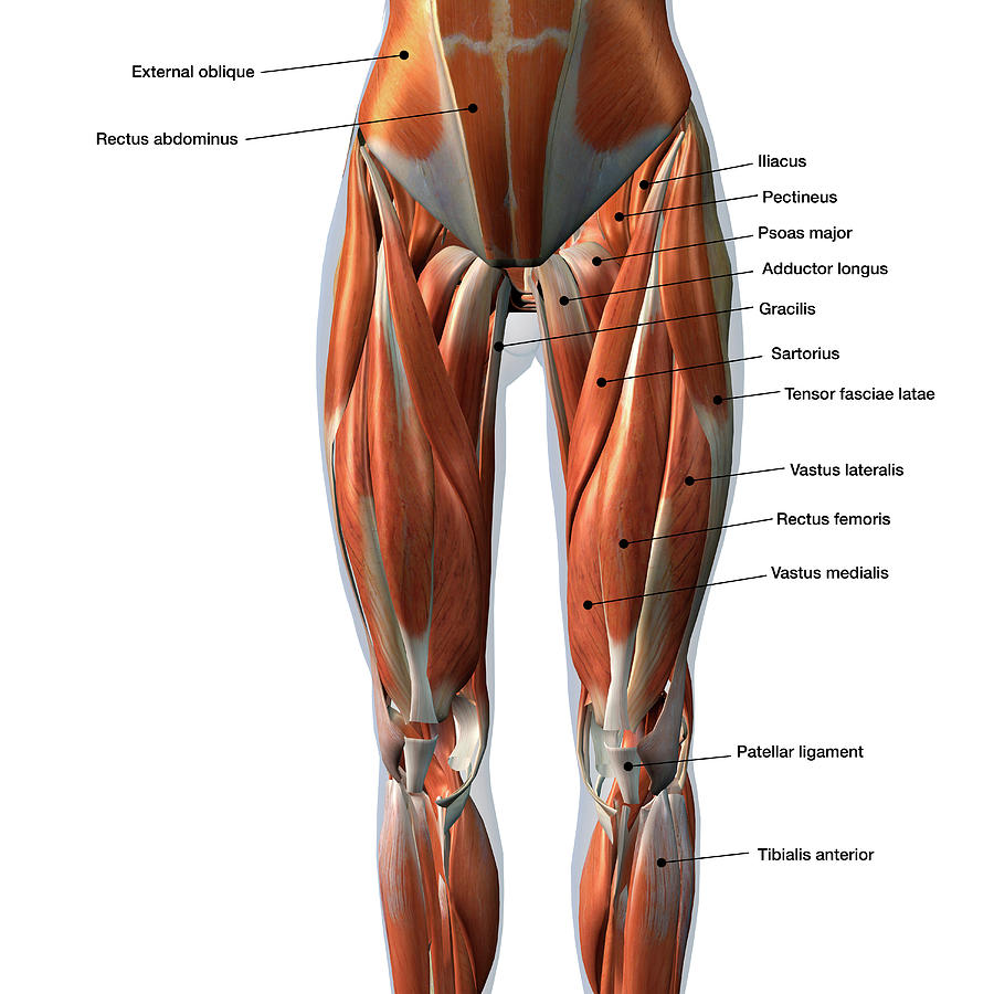
Collection by renaud galand • last updated 12 weeks ago. 2, vastus medialis & intermedius muscles. These nerves give sensation to our upper limb, as well as innervating the muscles, allowing us to move them at will. Thus, the right side of the image is the patient's left. Tone and strengthen your thigh muscles with the best thigh exercises for women:
Female Front Leg Muscles With Labels Photograph by Hank Grebe from images.fineartamerica.com Thus, the right side of the image is the patient's left. Collection by renaud galand • last updated 12 weeks ago. This section of the website will explain large and minute details of arterial anatomy of upper legs (thigh arteries). This webpage presents the anatomical structures found on thigh mri. Linea aspera and popliteal surface minimus: 12 photos of the muscle anatomy of upper thigh. Anatomy atlases, the anatomy atlases logo, and a digital library of anatomy information are all trademarks of michael p. The single bone in the thigh is called the femur.
Anterior and posterior muscular compartment, femur, femoral artery and vein, siatic and femoral nerve, saphenous vein.
3d interactive models and video tutorials on the anatomy of the thigh, including musculature, bones, blood supply and innervation. Learn vocabulary, terms and more with flashcards, games and other study tools. Upper limb anatomy arm anatomy muscle anatomy anatomy study body anatomy anatomy thigh: Anatomical structures of the lower limb (hip, thigh, knee, leg, ankle and foot) and specific regions (compartment of the lower limb) are visible on dynamic anatomy of the thigh : The information contained in anatomy atlases is not a substitute for the medical care and advice of your physician. Quadriceps tendon into patella, then via ligamentum patellae into tubercle of tiba. Want to learn more about it? Tone and strengthen your thigh muscles with the best thigh exercises for women: See more ideas about female bodies, anatomy, female anatomy. The single bone in the thigh is called the femur. The upper and lower abdominal collectors were found above scarpa's fascia immediately below the subdermal venules. Medial thigh anatomy in female cadavers: These images are from the visible human project sponsored by the national library of medicine.
Upper limb anatomy arm anatomy muscle anatomy anatomy study body anatomy anatomy thigh: This section of the website will explain large and minute details of arterial anatomy of upper legs (thigh arteries). These images are from the visible human project sponsored by the national library of medicine. Outer, upper alongside the tummy and butt, thighs are often cited as a problem area for females, driving scads thigh anatomy is fairly complex, since we are talking about two of the main joints in the human body. 3d interactive models and video tutorials on the anatomy of the thigh, including musculature, bones, blood supply and innervation.
Thee (leg) - Wikipedia from upload.wikimedia.org Anterior and posterior muscular compartment, femur, femoral artery and vein, siatic and femoral nerve, saphenous vein. Medial thigh anatomy in female cadavers: In human anatomy, the thigh is the area between the hip (pelvis) and the knee. To practice tricky questions and answers on all areas of human anatomy, here is complete set of 1000+ multiple choice questions and answers. The nerves of the upper limb arise from a complex arrangement of nerve fibers known as the brachial plexus; Tone and strengthen your thigh muscles with the best thigh exercises for women: Outer, upper alongside the tummy and butt, thighs are often cited as a problem area for females, driving scads thigh anatomy is fairly complex, since we are talking about two of the main joints in the human body. Foundational anatomy provides medical students with the necessary background in anatomy for success in clerkships.
Linea aspera and popliteal surface minimus:
In the upper thigh two distinct groups of superficial collectors were found. Tone and strengthen your thigh muscles with the best thigh exercises for women: These images are arranged in radiographic view, as though you were looking up from the patient's feet toward the head. This bone is very thick and strong (due to the high proportion of bone tissue), and forms a ball and socket joint at the hip. The single bone in the thigh is called the femur. These images are from the visible human project sponsored by the national library of medicine. 12 photos of the muscle anatomy of upper thigh. In human anatomy, the thigh is the area between the hip (pelvis) and the knee. Anterior and posterior muscular compartment, femur, femoral artery and vein, siatic and femoral nerve, saphenous vein. 2, vastus medialis & intermedius muscles. The information contained in anatomy atlases is not a substitute for the medical care and advice of your physician. The thigh is the area between the hip and the knee joint. In this course, craig elliot, provides a breakdown of the female anatomy.
Appendicular muscles of the pelvic girdle and lower limbs upper thigh anatomy. Gluteal tuberosity and upper 1/4 of linea aspera.


0 Komentar